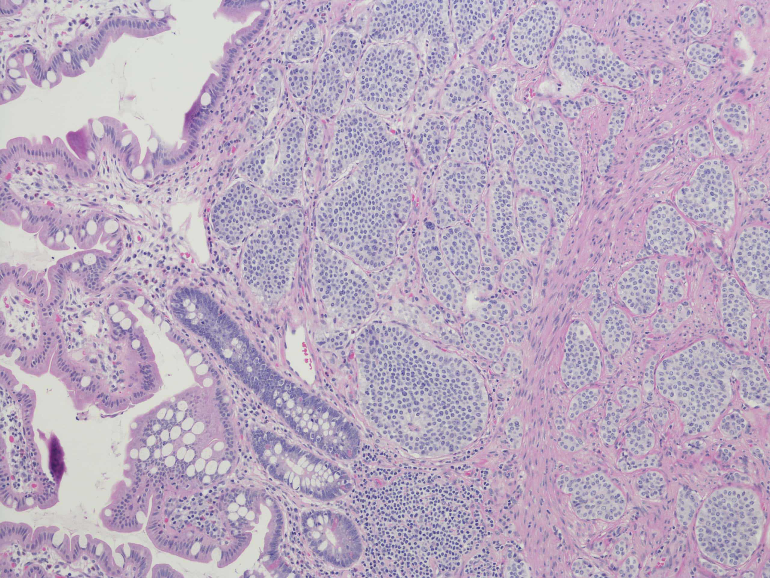
Neuroendocrine Tumour Imaging and Molecular Radiotherapy
Neuroendocrine tumours are a diverse group of relatively rare cancers, with a reported incidence of 9 cases per 100,000 people in 2015.
Recently there have been major advances in their diagnosis, staging and treatment. The possibility of multiple options to investigate and treat a diverse range of rare tumours represents a challenge for all members of the MDT, not least the radiologist.
The course aims to demystify this disease group and help radiologists achieve optimal management of their patients at the MDT.
It is expected that candidates have prior knowledge of FDG PET-CT imaging before undertaking the tutorials.
Learning Objectives
- Discuss the concept of somatostatin receptor imaging, normal distribution and variants of uptake, as well as some interpretation pitfalls.
- Review and illustrate the clinical indications for performing 68 Ga DOTA-SSA PET-CT, including the staging of well-differentiated NETs, the detection of occult NET primary tumours, and the use of 68 Ga DOTA-SSA PET-CT in evaluating recurrence.
- Discuss the role of 18FDG PET-CT in high- and low-grade neuroendocrine tumours and the potential role of 18F-FDOPA PET-CT.
- Explain the concept of molecular radiotherapy, review patient selection criteria and discuss the evidence for the use of PRRT in NET.
-
Ami Chander
Dr Chander is dually accredited in Diagnostic Radiology and Nuclear Medicine – and has a specialist interest in lung malignancy.
-
Tom Westwood
Dr Westwood is an Oncology Radiologist with a specialist interest in nuclear medicine and molecular Imaging. His experience includes a range of PET tracers including 18 FDG, 68Ga-DOTANOC, 18F-Choline and 18F-DOPA.

